
Corneal Cross-Linking (CXL) for Keratoconus
Corneal collagen cross-linking · Prof. Dr. Fırat Helvacıoğlu, FEBOS-CR · Veni Vidi Eye Clinic, Caddebostan – Kadıköy, Istanbul
Corneal cross-linking (CXL) is a treatment that halts the progression of keratoconus by strengthening the collagen fibres of the cornea with riboflavin drops and controlled UVA light. Surgeons perform it under topical anaesthesia, and it takes 30–60 minutes per eye. Its aim is not to correct vision but to halt the disease. In young patients, studies have shown that it reduces the risk of progression by about 90%.
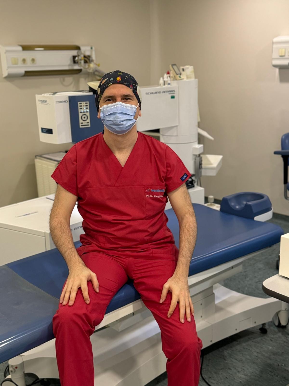
What is corneal cross-linking (CXL)?
Corneal cross-linking (CXL) strengthens the cornea by creating new bonds between its collagen fibres using riboflavin (vitamin B2) drops and controlled ultraviolet-A (UVA) light. As a result, the strengthened cornea becomes more resistant, and keratoconus stops progressing.
The main aim of CXL is not to correct vision but to halt disease progression. Some patients experience slight corneal flattening and improved vision, but patients usually still correct the existing prescription with glasses, lenses or additional treatments.
Who needs corneal cross-linking?
- Progressive keratoconus: Increasing corneal steepness, thinning or a significant change in prescription on consecutive measurements.
- Children and young people: Because the disease can progress rapidly at these ages, we recommend early treatment once we detect progression.
- Ectasia after laser or corneal surgery: To halt corneal weakening.
- Adequate corneal thickness: Usually at least 400 microns for the standard method; we use special protocols for thinner corneas.
However, CXL may not suit very advanced cases with corneal scarring, or patients with active infection or severe ocular surface disease.
What happens during corneal cross-linking (CXL)?
- Anaesthesia: Drops numb the eye; no pain is felt during the procedure, although stinging and light sensitivity may occur in the first days.
- Surface layer: In the standard (epi-off) method, the surgeon gently removes the surface layer (epithelium) so that riboflavin can penetrate the cornea.
- Riboflavin: We apply riboflavin drops at set intervals until riboflavin saturates the cornea.
- UVA light: We irradiate the cornea with a controlled dose of UVA light. In accelerated protocols, the irradiation time is shorter.
- Protective lens: We place a clear bandage contact lens on the eye until the surface layer has healed.
Overall, the whole procedure takes about 30–60 minutes per eye, and the patient goes home the same day.
Corneal cross-linking (CXL) step by step: illustration
Strengthening the cornea with riboflavin drops and UVA light (epi-off method).
- Removing the epitheliumDrops numb the eye. In the standard (epi-off) method, the surgeon gently removes the surface layer (epithelium) so that riboflavin can penetrate the cornea.
- Riboflavin dropsWe apply riboflavin (vitamin B2) drops at set intervals until riboflavin saturates the corneal tissue.
- UVA lightWe irradiate the cornea with a controlled dose of UVA light. In the classic protocol this takes 30 minutes; accelerated protocols are shorter.
- The strengthened corneaNew bonds form between the collagen fibres; the cornea stiffens and keratoconus stops progressing. You wear a protective lens until the epithelium heals.
Standard versus accelerated CXL
In the classic “Dresden” protocol, UVA light is applied for 30 minutes; in accelerated protocols, higher-intensity light shortens the time. Meta-analyses of randomised trials have shown that both methods halt disease progression similarly; the standard protocol may achieve slightly more corneal flattening and a deeper effect, while the accelerated protocol causes less corneal thinning. Finally, the surgeon chooses the protocol according to corneal thickness and disease stage.
Recovery after corneal cross-linking
| Time | What to expect |
|---|---|
| Days 1–3 | Grittiness, watering, light sensitivity; controlled with painkillers and drops |
| Days 3–5 | The surface layer closes and we remove the bandage lens |
| Week 1 – month 1 | Vision may fluctuate; most patients return to desk work within 1 week |
| Months 3–12 | The cornea stabilises; we decide about contact lens wear at a follow-up visit |
You may notice mild corneal haze in the first months; it usually fades over time. Serious complications such as infection are rare, which is why regular use of drops and attending follow-up visits are important.
Success of corneal cross-linking and the evidence
Randomised trials show that corneal cross-linking is the core treatment to halt progression in keratoconus. For instance, in the KERALINK trial, the likelihood of progression in young patients treated with CXL was about 90% lower than with standard monitoring. Meta-analyses in paediatric patients have also reported that all CXL methods slow progression for at least one year. In a small number of patients, progression may continue despite treatment; in that case, we can repeat CXL.
Cost and international patients: We determine the cost after the examination. Because the surface layer takes 3–5 days to heal and we remove the bandage lens at the follow-up visit, we recommend that international patients plan a stay of about one week in Istanbul. Finally, send us your corneal topography/tomography via WhatsApp for a preliminary assessment.
Related treatments: Combined CXL + No Touch · CAIRS and intracorneal ring segments (ICRS) · Keratoconus treatment
Frequently asked questions
The aim of CXL is not to correct vision but to halt the progression of keratoconus. Some patients experience slight improvement; patients correct the remaining prescription with glasses, lenses or combined treatments.
In most patients a single treatment is sufficient and its effect lasts for many years. However, if progression continues at follow-up, we can repeat the treatment.
Once the surface layer has healed and the cornea has stabilised; we usually decide at a follow-up visit within 1–3 months.
The epi-on method causes less pain and heals faster, but studies generally find it less effective at halting progression than the standard epi-off method. We make the choice according to the patient’s condition.
Keratoconus can progress faster in children. So when we detect progression, early CXL reduces the future risk of vision loss and corneal transplantation.
References
- Larkin DFP et al. Effect of corneal cross-linking versus standard care on keratoconus progression in young patients: the KERALINK randomized controlled trial. Ophthalmology. 2021;128(11):1516–1526. doi:10.1016/j.ophtha.2021.04.019
- Shajari M et al. Comparison of standard and accelerated corneal cross-linking for the treatment of keratoconus: a meta-analysis. Acta Ophthalmol. 2019;97(1):e22–e35. doi:10.1111/aos.13814
- Kobashi H, Tsubota K. Accelerated versus standard corneal cross-linking for progressive keratoconus: a meta-analysis of randomized controlled trials. Cornea. 2020;39(2):172–180. doi:10.1097/ICO.0000000000002092
- Kobashi H et al. Corneal cross-linking for paediatric keratoconus: a systematic review and meta-analysis. J Clin Med. 2021;10(12):2626. doi:10.3390/jcm10122626
- Gomes JA et al. Global consensus on keratoconus and ectatic diseases. Cornea. 2015;34(4):359–369. doi:10.1097/ICO.0000000000000408
This page offers patient information and does not replace an examination or medical assessment. We determine the right treatment for you after an eye examination and measurements.
Book a cross-linking assessment
Prof. Dr. Fırat Helvacıoğlu · Veni Vidi Eye Clinic, Bağdat Caddesi No: 257, Caddebostan – Kadıköy, Istanbul, Türkiye