
Medical Retina
Diabetic retinopathy · Macular degeneration · Retinitis pigmentosa · Prof. Dr. Fırat Helvacıoğlu, FEBOS-CR · Veni Vidi Eye Clinic, Caddebostan – Kadıköy, Istanbul
Medical retina is the management of retinal diseases such as diabetic retinopathy, macular degeneration and retinitis pigmentosa with intravitreal injections (anti-VEGF), laser and medical treatment. Because these diseases often progress silently, early diagnosis with regular dilated fundus examination and OCT is the key to preserving vision.
What are retinal diseases?
Medical retina is the field of ophthalmology that treats diseases of the retina — the light-sensitive layer at the back of the eye — and the macula with medication, laser and intravitreal injections. The retina lines the inside of the eye like a film and converts light into nerve signals; the macula at its centre is responsible for reading, recognising faces and sharp vision.
Retinal diseases often cause no symptoms in their early stages. Regular dilated fundus examination is therefore the most effective way to prevent vision loss, particularly in people with diabetes, those over 50 and those with a family history of retinal disease.
Main retinal diseases we treat
| Disease | What happens? | Main treatment |
|---|---|---|
| Diabetic retinopathy | Diabetes damages the retinal blood vessels, causing leakage, haemorrhage and new vessel growth | Blood sugar control, intravitreal injections (anti-VEGF), retinal laser, vitrectomy if needed |
| Macular degeneration (AMD) | Age-related damage to the macula; central vision deteriorates | Intravitreal injections for the wet form; monitoring, nutritional supplements and risk factor control for the dry form |
| Night blindness (retinitis pigmentosa) | Inherited loss of rod and cone cells; night and peripheral vision decline | Treatment of complications (macular oedema, cataract), genetic counselling, gene therapy in eligible patients |
Examination and imaging of retinal diseases
- Dilated fundus examination: First, we dilate the pupil with drops and examine the retina and optic nerve in detail.
- Optical coherence tomography (OCT): A non-contact scan that shows the layers of the retina at micron level; it is the key tool for detecting macular oedema and fluid and for monitoring treatment.
- Fundus photography and angiography: When needed, in addition, dye-based imaging shows leaking vessels and new vessels.
- Visual field and colour vision tests: Used particularly to monitor the stage of inherited retinal diseases.
Treatments for retinal diseases
- Intravitreal injections (anti-VEGF): We inject drugs that stop abnormal vessel growth and leakage into the eye; this is the standard treatment for wet macular degeneration and diabetic macular oedema.
- Intravitreal steroids: Used in selected cases of macular oedema.
- Retinal laser (panretinal photocoagulation): Reduces new vessel growth and the risk of haemorrhage in proliferative diabetic retinopathy.
- Drops and oral medication: For example, for cystoid macular oedema in retinitis pigmentosa.
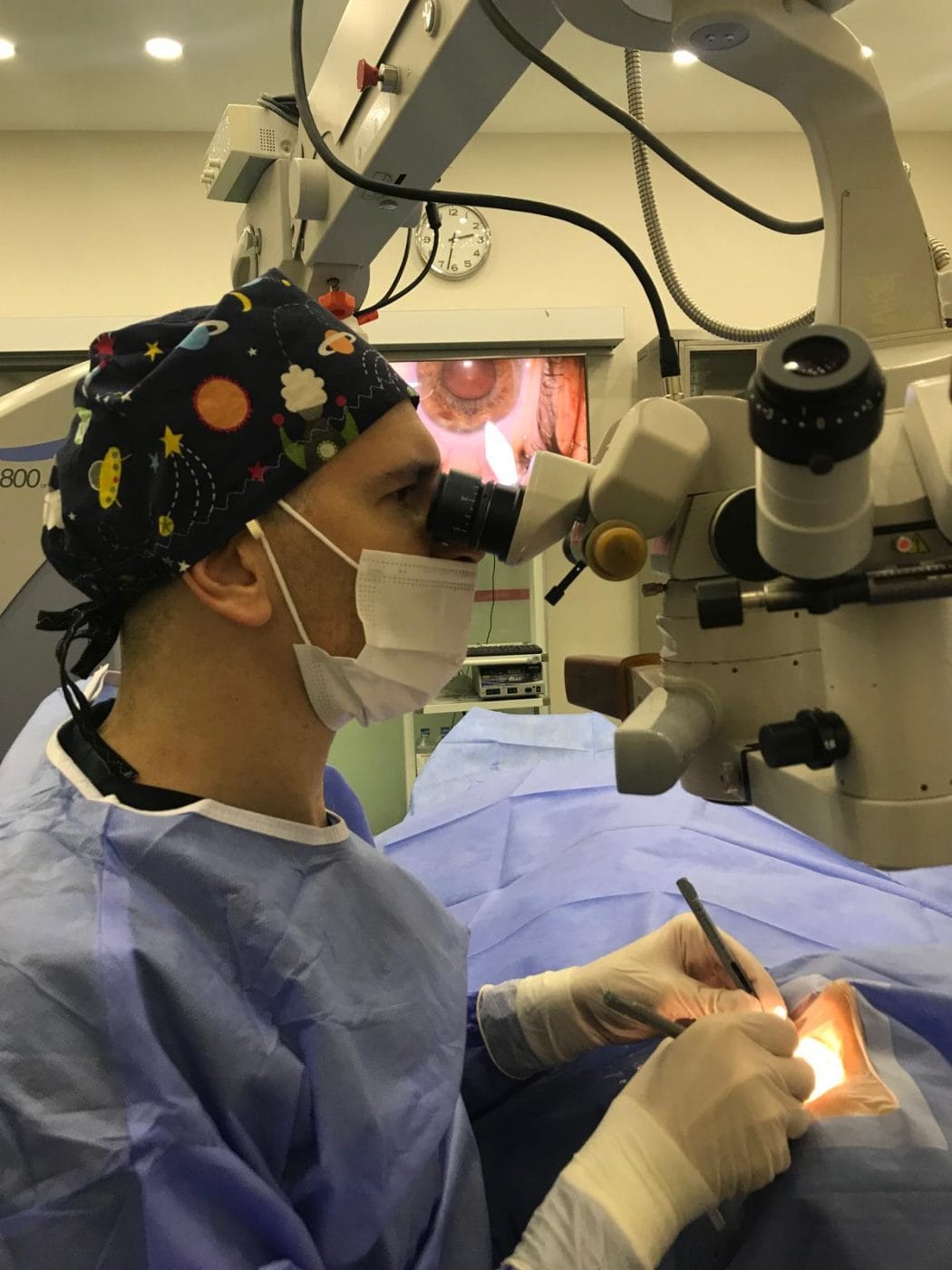
How is an intravitreal injection performed? We carry out the procedure under sterile operating conditions with topical anaesthesia and takes a few minutes. Then we clean the eye and surrounding area with an antiseptic solution, hold the eyelids open with a speculum, and inject the medication into the eye through the white of the eye with a very fine needle. Most patients feel only slight pressure. You may use antibiotic drops for a few days afterwards; if pain, increasing redness or reduced vision occur, seek medical attention immediately. Serious side effects such as infection (endophthalmitis) are rare.
Prof. Dr. Fırat Helvacıoğlu’s work in the retina field
Research
With Prof. Dr. Zafer Kapran, Prof. Dr. Fırat Helvacıoğlu wrote the chapter “Anti-VEGF therapy in age-related macular degeneration” in a special issue of the journal Retina-Vitreus (2015). He published the “Helvacıoğlu reproducibility index”, an algorithm named after him that evaluates the effect of misalignment on retinal nerve fibre layer measurements with spectral-domain OCT, in the International Journal of Ophthalmology (2015). His other retinal work includes OCT findings of macular folds in nanophthalmos and the formation of eccentric macular holes after vitrectomy.
Frequently asked questions
Doctors can treat many of them or slow their progression. In wet macular degeneration and diabetic macular oedema, intravitreal injections can preserve and even improve vision. Early diagnosis is the most important factor determining the outcome.
We perform the procedure with anaesthetic drops; most patients feel only slight pressure. It takes a few minutes and you go home the same day.
For people with diabetes without retinopathy, we generally recommend an annual dilated fundus examination; if we detect retinopathy, we shorten the interval according to its stage. Also, pregnancy requires more frequent examinations.
The pupil-dilating drops cause blurred vision and light sensitivity for a few hours, so we advise you not to drive during this time.
References
- Teo ZL et al. Global prevalence of diabetic retinopathy and projection of burden through 2045: systematic review and meta-analysis. Ophthalmology. 2021;128(11):1580–1591. doi:10.1016/j.ophtha.2021.04.027
- Wong WL et al. Global prevalence of age-related macular degeneration and disease burden projection for 2020 and 2040: a systematic review and meta-analysis. Lancet Glob Health. 2014;2(2):e106–e116. doi:10.1016/S2214-109X(13)70145-1
- Virgili G et al. Anti-vascular endothelial growth factor for diabetic macular oedema: a network meta-analysis. Cochrane Database Syst Rev. 2023;6(6):CD007419. doi:10.1002/14651858.CD007419.pub7
- Solomon SD et al. Anti-vascular endothelial growth factor for neovascular age-related macular degeneration. Cochrane Database Syst Rev. 2019;3(3):CD005139. doi:10.1002/14651858.CD005139.pub4
- Hartong DT et al. Retinitis pigmentosa. Lancet. 2006;368(9549):1795–1809. doi:10.1016/S0140-6736(06)69740-7
- Kapran Z, Helvacıoğlu F. Anti-VEGF therapy in age-related macular degeneration. Retina-Vitreus. 2015;Special issue:233–243 (in Turkish).
- Helvacıoğlu F, Uyar OM, Şencan S, Tunç Z, Kapran Z. Helvacioglu reproducibility index: a new algorithm to evaluate the effects of misalignments on the measurements of retinal nerve fiber layer by spectral-domain OCT. Int J Ophthalmol. 2015;8(5). doi:10.3980/j.issn.2222-3959.2015.05.28
This page is intended for patient information and does not replace an examination or medical assessment. Therefore, we determine the right treatment for you after an eye examination and measurements.
Book a retinal examination and OCT assessment
Prof. Dr. Fırat Helvacıoğlu · Veni Vidi Eye Clinic, Bağdat Caddesi No: 257, Caddebostan – Kadıköy, Istanbul, Türkiye