
Glaucoma Treatment
Open-angle and angle-closure glaucoma · SLT laser · Glaucoma surgery · Prof. Dr. Fırat Helvacıoğlu, FEBOS-CR · Veni Vidi Eye Clinic, Caddebostan – Kadıköy, Istanbul
Glaucoma is a disease that damages the optic nerve, most often due to raised intraocular pressure, and vision that is lost does not return. Its most common type causes no symptoms for years; doctors make the diagnosis by measuring eye pressure, OCT and visual field testing. Glaucoma treatment preserves vision by lowering the pressure with drops, SLT laser, laser iridotomy or surgery.
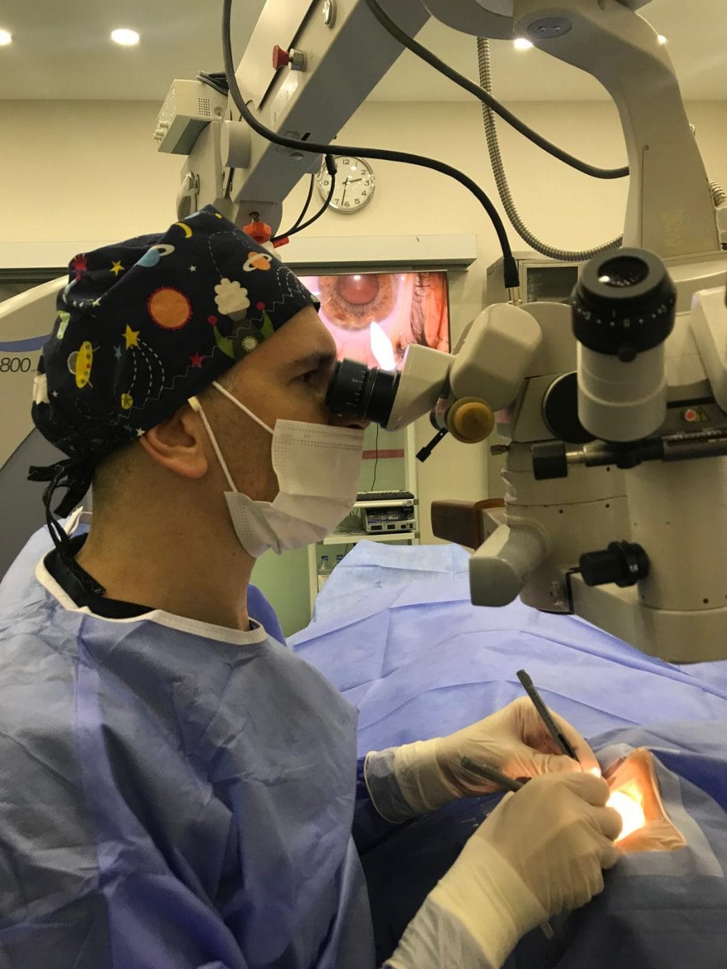
What is glaucoma?
Glaucoma is a group of diseases that progressively damage the optic nerve, leading to permanent vision loss if left untreated. The most important risk factor is high intraocular pressure; however, glaucoma can also develop at normal pressure. Vision lost to glaucoma does not return, so early diagnosis and regular treatment are vital.
Glaucoma is the leading cause of irreversible blindness worldwide. According to a meta-analysis, about 3.5% of people aged 40–80 have glaucoma, and the meta-analysis projects that the number of people with glaucoma worldwide will exceed 111 million by 2040. In addition, a significant proportion of patients are unaware of their disease.
How does glaucoma develop? Watch the animation
How does glaucoma develop? Interactive animation
See how the fluid inside the eye (aqueous humour) flows, why the pressure rises, and how damage to the optic nerve narrows the visual field.
Normal intraocular pressure is generally between 10 and 21 mmHg; however, glaucoma can also develop at normal pressure. Illustrative animation; proportions have been simplified for clarity. © Prof. Dr. Fırat Helvacıoğlu
Types of glaucoma
| Type | Features | Symptoms |
|---|---|---|
| Primary open-angle glaucoma | The most common type; the angle is open, but outflow through the trabecular meshwork is reduced | No symptoms for years; the visual field narrows insidiously from the edges |
| Angle-closure glaucoma | The angle between the iris and the cornea narrows or closes; more common in hyperopic and small eyes and as the lens thickens with age | In an acute attack: severe pain, redness, blurred vision, halos around lights, nausea and vomiting |
| Normal-tension glaucoma | Nerve damage develops even though the pressure is within normal limits | No symptoms; diagnosed by examining the nerve and visual field |
| Secondary glaucomas | Due to pseudoexfoliation, pigment dispersion, uveitis, diabetes (neovascular), corticosteroid use or trauma | Varies with the cause |
| Congenital glaucoma | Developmental abnormality of the angle in infants | Large, cloudy cornea, watering, avoidance of light |
Who is at risk?
- Everyone over 40 (the risk increases with age)
- People with a family history of glaucoma
- People with raised eye pressure (ocular hypertension)
- Highly myopic people (open-angle) and hyperopic people (angle-closure)
- People with diabetes, high or very low blood pressure, migraine or sleep apnoea
- Long-term users of corticosteroid drops, sprays or medication
- People with thin corneas; previous eye injury
If you are in a risk group, you should have regular eye examinations even if you have no symptoms. We recommend a comprehensive eye examination for everyone from the age of 40.
How do we diagnose glaucoma before glaucoma treatment?
- Eye pressure measurement (tonometry): We also assess it together with corneal thickness measurement (pachymetry); in thin corneas the pressure may read lower than it really is.
- Angle examination (gonioscopy) and anterior segment OCT: Shows whether the angle is open or narrow.
- Optic nerve examination and OCT: In addition, OCT measures the nerve fibre layer and ganglion cell complex at micron level, so it can detect damage before it affects the visual field.
- Visual field test (perimetry): Shows the effect of the disease on vision and whether it is progressing.
Prof. Dr. Fırat Helvacıoğlu’s work on glaucoma imaging
Prof. Dr. Fırat Helvacıoğlu developed the “Helvacıoğlu reproducibility index”, an algorithm named after him that evaluates how scan misalignment affects OCT retinal nerve fibre layer measurements, and published it in the International Journal of Ophthalmology (2015). His study of the relationship between the nerve fibre layer and the ganglion cell complex in glaucomatous and healthy eyes appeared in the Turkish Journal of Ophthalmology (Türk Oftalmoloji Gazetesi) (2010). He has also presented at national congresses on anterior segment OCT analysis of the angle after YAG laser iridotomy in angle-closure glaucoma and on SLT in glaucoma.
Glaucoma treatment options
The aim of treatment is to lower the eye pressure to a “target pressure” that halts nerve damage in the individual patient. In the Early Manifest Glaucoma Trial, lowering the pressure by an average of 25% reduced the rate of progression over 6 years from 62% to 45%.
| Treatment | How does it work? | Suitable for |
|---|---|---|
| Eye drops | Reduce fluid production or increase outflow | First choice in most patients; requires regular, lifelong use |
| Selective laser trabeculoplasty (SLT) | Stimulates the trabecular meshwork with low-energy laser to increase outflow; a few-minute outpatient procedure | Open-angle glaucoma and ocular hypertension, as first-line treatment or in addition to drops |
| YAG laser iridotomy | Creates a small opening in the iris so that fluid can pass from the posterior to the anterior chamber, preventing angle closure | Narrow angles, angle-closure glaucoma and acute attacks |
| Lens (cataract/refractive lens) surgery | Removing the thickened natural lens widens the angle | Selected patients with angle-closure glaucoma |
| Glaucoma surgery (trabeculectomy, tube/valve, MIGS) | Creates a new outflow pathway for the fluid | Patients whose pressure cannot be controlled, or whose disease progresses, despite drops and laser |
In the LiGHT trial of 718 patients, 74% of those treated with SLT as first-line therapy remained at target pressure without drops for three years; six-year results showed less disease progression (19.6% vs 26.8%) and less need for glaucoma surgery in the SLT group. The EAGLE trial showed that early clear lens extraction in angle-closure glaucoma provides lower pressure and better quality of life than laser and drop treatment.
Prof. Dr. Fırat Helvacıoğlu plans glaucoma treatment — drop regimens, SLT and YAG laser iridotomy, glaucoma surgery and combined cataract–glaucoma approaches — according to the type and stage of each patient’s disease.
Acute glaucoma attack: emergency symptoms
Sudden severe eye pain and headache, eye redness, blurred vision, coloured halos around lights, nausea and vomiting may indicate an acute angle-closure attack. As it can cause permanent vision loss within hours, see an ophthalmologist or go to the emergency department without delay.
Glaucoma treatment in daily life: what to keep in mind
- Use your drops at the same times every day without missing doses; if you use more than one drop, wait at least 5 minutes between them.
- Gently pressing on the inner corner of the eye for 1–2 minutes after instilling a drop reduces absorption of the medication into the bloodstream.
- Do not miss your follow-up appointments and visual field tests; the disease can progress without symptoms.
- Always tell other doctors that you receive glaucoma treatment, because some medications (for example corticosteroids and some cold remedies) can affect eye pressure.
- Make sure your first-degree relatives also have eye examinations.
Frequently asked questions about glaucoma treatment
No. Nerve fibres damaged by glaucoma do not regenerate. The aim of treatment is to preserve the remaining vision, which is why early diagnosis is so important.
Not always. Doctors call raised pressure without nerve damage ocular hypertension; these people are at risk of glaucoma, so we monitor them and start treatment when necessary.
In most patients, yes. However, SLT laser or surgery may reduce or eliminate the need for drops.
SLT is performed with anaesthetic drops and takes a few minutes; no pain is felt during the procedure, although mild pressure or stinging may occur. You can return to your daily activities the same day.
The risk is markedly higher in people with a family history of glaucoma. Therefore, we advise people whose first-degree relatives have glaucoma to have an examination even before the age of 40.
Particularly in narrow-angle and angle-closure glaucoma, removing the lens can widen the angle and lower the pressure. However, examination and angle measurements decide suitability.
References
- Tham YC et al. Global prevalence of glaucoma and projections of glaucoma burden through 2040: a systematic review and meta-analysis. Ophthalmology. 2014;121(11):2081–2090. doi:10.1016/j.ophtha.2014.05.013
- Heijl A et al. Reduction of intraocular pressure and glaucoma progression: results from the Early Manifest Glaucoma Trial. Arch Ophthalmol. 2002;120(10):1268–1279. doi:10.1001/archopht.120.10.1268
- Gazzard G et al. Selective laser trabeculoplasty versus eye drops for first-line treatment of ocular hypertension and glaucoma (LiGHT): a multicentre randomised controlled trial. Lancet. 2019;393(10180):1505–1516. doi:10.1016/S0140-6736(18)32213-X
- Gazzard G et al. Laser in Glaucoma and Ocular Hypertension (LiGHT) trial: six-year results of primary selective laser trabeculoplasty versus eye drops for the treatment of glaucoma and ocular hypertension. Ophthalmology. 2023;130(2):139–151. doi:10.1016/j.ophtha.2022.09.009
- He M et al. Laser peripheral iridotomy for the prevention of angle closure: a single-centre, randomised controlled trial. Lancet. 2019;393(10181):1609–1618. doi:10.1016/S0140-6736(18)32607-2
- Azuara-Blanco A et al. Effectiveness of early lens extraction for the treatment of primary angle-closure glaucoma (EAGLE): a randomised controlled trial. Lancet. 2016;388(10052):1389–1397. doi:10.1016/S0140-6736(16)30956-4
- Helvacıoğlu F, Uyar OM, Şencan S, Tunç Z, Kapran Z. Helvacioglu reproducibility index: a new algorithm to evaluate the effects of misalignments on the measurements of retinal nerve fiber layer by spectral-domain OCT. Int J Ophthalmol. 2015;8(5). doi:10.3980/j.issn.2222-3959.2015.05.28
- Korkmaz B, Yiğit U, Ağaçhan A, Helvacıoğlu F, Bilen H, Tuğcu B. Evaluation of the relationship between retinal nerve fibre layer and ganglion cell complex measured by optical coherence tomography in glaucomatous and normal eyes. Türk Oftalmoloji Gazetesi. 2010;40:338–342 (in Turkish).
- Helvacıoğlu F, Akıngöl Z, Şencan S, Tunç Z. Selektif Laser Trabeküloplasti Sonrası Gözlenen Erken Dönem Göz İçi Basıncı Artışı ve Risk Faktörleri. Türk Oftalmoloji Dergisi. 2014;44(5):365-369. doi:10.4274/tjo.69077
This page offers patient information and does not replace an examination or medical assessment. Therefore, we determine the treatment that is right for you after an eye examination and measurements.
Glaucoma screening: eye pressure, OCT and visual field
Prof. Dr. Fırat Helvacıoğlu · Veni Vidi Eye Clinic, Bağdat Caddesi No: 257, Caddebostan – Kadıköy, Istanbul, Türkiye
Related pages
- Comprehensive Eye ExaminationWho needs an eye examination and how often, what a comprehensive eye exam involves, and which symptoms need urgent care. Veni Vidi Eye Clinic, Istanbul.
- Diabetic RetinopathyDiabetic retinopathy and diabetic macular oedema: stages, screening intervals, anti-VEGF injections, retinal laser and prevention. Istanbul.
- Cataract SurgeryPhaco cataract surgery with monofocal, toric, EDOF or trifocal lenses by FEBOS-CR surgeon Prof. Dr. Fırat Helvacıoğlu in Kadıköy, Istanbul.
- Eye Diseases and TreatmentsGlaucoma, dry eye, blepharitis, pterygium, corneal diseases, tear duct obstruction, strabismus and eyelid surgery in Istanbul. Diagnosis and treatment.