
Corneal Diseases and Treatments
Keratitis · Fuchs dystrophy · Keratoconus · Corneal transplantation · Prof. Dr. Fırat Helvacıoğlu, FEBOS-CR · Veni Vidi Eye Clinic, Caddebostan – Kadıköy, Istanbul
The cornea is the clear front layer of the eye; corneal diseases such as infection (keratitis), keratoconus, Fuchs endothelial dystrophy, injuries and scars reduce vision by disturbing its clarity or shape. Depending on the disease, treatment includes drops, cross-linking, specialty lenses, laser or corneal transplantation. Therefore, pain and redness while wearing contact lenses need urgent assessment.
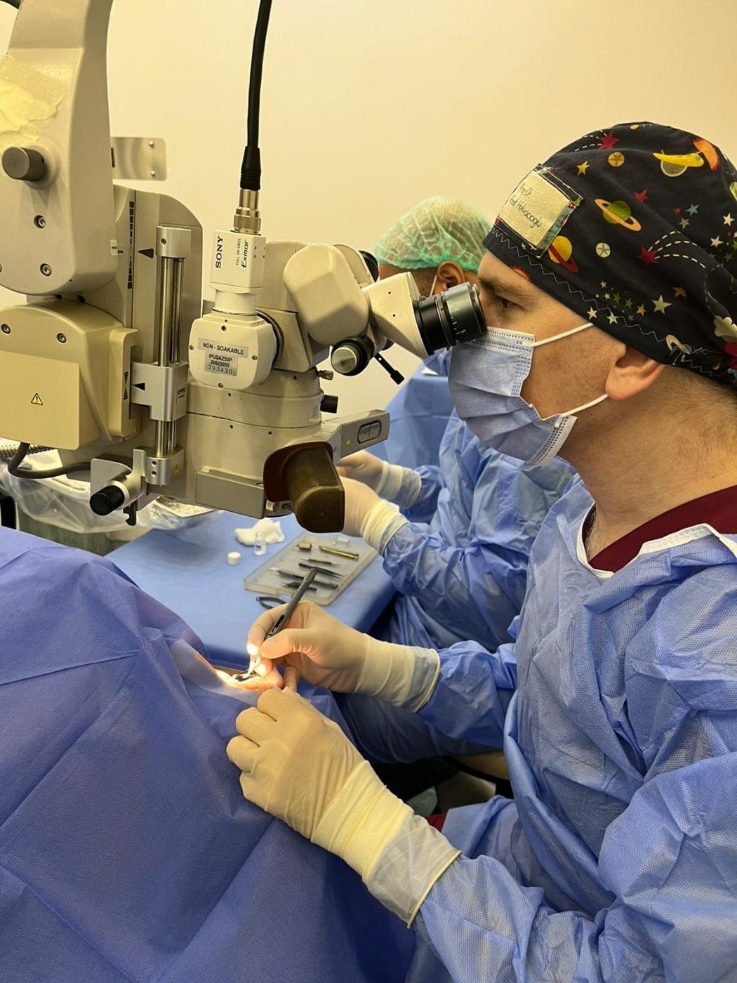
What is the cornea, and which corneal diseases affect it?
The cornea is the clear, dome-shaped layer at the very front of the eye; it provides about two thirds of the eye’s focusing power. As a result, any disease that disturbs its clarity or shape affects vision. The main corneal diseases are:
| Disease | Features | Treatment |
|---|---|---|
| Keratitis and corneal ulcer | Infection caused by bacteria, fungi, viruses (herpes) or Acanthamoeba; the risk is higher in contact lens wearers | Intensive antibiotic/antiviral/antifungal drops; surgery in severe cases |
| Keratoconus | Thinning and forward bulging of the cornea into a cone | Cross-linking, corneal ring segments, specialty lenses, transplantation |
| Fuchs endothelial dystrophy | Loss of the pump cells on the inner surface of the cornea; blurred vision in the morning, oedema | Drops; endothelial transplantation (DMEK) in advanced stages |
| Epithelial basement membrane dystrophy and recurrent erosion | Poor adhesion of the surface layer; sudden pain on waking | Ointments, bandage lens, surface treatment (PTK) |
| Corneal scars and injuries | Permanent opacity after infection or trauma | Rigid lens, laser (PTK), corneal transplantation |
| Dry eye and ocular surface diseases | Tear film disorders, limbal stem cell deficiency | Dry eye treatment, amniotic membrane, stem cell transplantation |
Contact lenses and corneal diseases: infection risk
Contact lens wear is one of the most important risk factors for corneal infection. In a study from Australia, the annual incidence of infection was about 2 per 10,000 wearers of daily-wear soft lenses, whereas it increased more than tenfold in those who slept in their lenses. In addition, sleeping in lenses, poor lens case hygiene, rinsing lenses with tap water and swimming with lenses all increase the risk.
If you develop eye pain, redness, light sensitivity or blurred vision while wearing contact lenses, remove the lens immediately and see an ophthalmologist the same day. This is because corneal infections can progress within hours.
Corneal transplantation (keratoplasty) for advanced corneal diseases
When corneal diseases permanently cloud the cornea or distort its shape too much to correct, the surgeon replaces the diseased tissue with healthy donor cornea. Today, whenever possible, the surgeon replaces only the diseased layer:
- Penetrating keratoplasty (PK): The surgeon replaces all layers of the cornea.
- Deep anterior lamellar keratoplasty (DALK): In keratoconus and superficial scars, the surgeon replaces the anterior layers and preserves the inner layer (endothelium); the risk of rejection is lower.
- Endothelial keratoplasty (DMEK/DSAEK): In endothelial diseases such as Fuchs dystrophy, the surgeon replaces only the inner layer. In a randomised trial, 66% of patients achieved 20/25 vision at 12 months after DMEK (33% after ultrathin DSAEK).
In 2012, surgeons performed about 185,000 corneal transplants in 116 countries worldwide; the main indications were Fuchs dystrophy (39%), keratoconus (27%) and post-infectious scars (20%). Moreover, according to the same study, there is only 1 donor cornea available for every 70 needed worldwide, which is why organ donation is vital.
Prof. Dr. Fırat Helvacıoğlu’s work on corneal diseases
Research and teaching
Prof. Dr. Fırat Helvacıoğlu is the author of the “Cornea and Ocular Surface Diseases” chapter of the book Güncel ve Özet Oftalmoloji (2018). In addition, he has published and presented at ESCRS/ASCRS on the establishment of the eye bank at Haseki Training and Research Hospital, rules for donor cornea preparation and use, donor serology, and the outcomes of keratoplasties performed with corneas from this bank. He published the culture results of vancomycin-treated donor rims in penetrating keratoplasty in Nobel Medicus. He published the results of mechanically implanted corneal ring segments in keratoconus and post-LASIK ectasia in the Indian Journal of Ophthalmology, and has served as a reviewer on corneal topics for Ophthalmology.
Frequently asked questions about corneal diseases
Corneal transplantation is one of the most successful tissue transplants; nevertheless, rejection can occur. If redness, pain, light sensitivity or reduced vision develop, seek care immediately; doctors can reverse most rejection episodes that they treat early.
It depends on the technique. After endothelial transplantation (DMEK), vision clears within weeks, whereas after full-thickness transplantation it may take months to reach final vision because of sutures and astigmatism.
Corneal donation is part of organ donation; eye colour, age or wearing glasses are usually not barriers to corneal donation. Also, you can register as an organ donor through the national organ donation registry in your country.
Yes, because the herpes virus lies dormant in the nerves, it can recur. For patients with frequent recurrences, doctors may recommend preventive antiviral treatment.
References
- Gain P et al. Global survey of corneal transplantation and eye banking. JAMA Ophthalmol. 2016;134(2):167–173. doi:10.1001/jamaophthalmol.2015.4776
- Stapleton F et al. The incidence of contact lens-related microbial keratitis in Australia. Ophthalmology. 2008;115(10):1655–1662. doi:10.1016/j.ophtha.2008.04.002
- Dunker SL et al. Descemet membrane endothelial keratoplasty versus ultrathin Descemet stripping automated endothelial keratoplasty: a multicenter randomized controlled clinical trial. Ophthalmology. 2020;127(9):1152–1159. doi:10.1016/j.ophtha.2020.02.029
- Ong Tone S et al. Fuchs endothelial corneal dystrophy: the vicious cycle of Fuchs pathogenesis. Prog Retin Eye Res. 2020;80:100863. doi:10.1016/j.preteyeres.2020.100863
- Helvacıoğlu F. Cornea and ocular surface diseases. In: Oruçoğlu F, ed. Güncel ve Özet Oftalmoloji. 2018:55–125 (in Turkish). ISBN 978-605-7993-19-9.
- Tunç Z, Helvacıoğlu F, Şencan S. Evaluation of intrastromal corneal ring segments for treatment of keratoconus with a mechanical implantation technique. Indian J Ophthalmol. 2013;61(5):218–225. doi:10.4103/0301-4738.109519
This page provides patient information and does not replace an examination or medical assessment. Finally, after an eye examination and measurements, we determine the treatment that is right for you.
Corneal examination and topography
Prof. Dr. Fırat Helvacıoğlu · Veni Vidi Eye Clinic, Bağdat Caddesi No: 257, Caddebostan – Kadıköy, Istanbul, Türkiye