
Cataract Surgery
Prof. Dr. Fırat Helvacıoğlu, FEBOS-CR · Cataract and Refractive Surgery · Veni Vidi Eye Clinic, Caddebostan – Kadıköy, Istanbul
Cataract surgery removes the clouded natural lens of the eye through a very small incision using ultrasound energy (phacoemulsification) and replaces it with a permanent, foldable intraocular lens (IOL). We perform the operation under topical (eye-drop) anaesthesia; it usually takes 10–20 minutes, and patients go home the same day. In most patients vision improves markedly within the first few days.
On this page
- What is a cataract?
- Symptoms and timing of surgery
- How is surgery performed, step by step?
- Intraocular lens options
- Monofocal lenses and monovision
- Technology and measurements
- Light phenomena after surgery (dysphotopsia)
- Risks and safety
- Secondary cataract and YAG laser
- Recovery
- Cost and international patients
- Why Prof. Dr. Fırat Helvacıoğlu?
- Frequently asked questions
What is a cataract?
A cataract is the loss of transparency, or clouding, of the eye’s natural crystalline lens. Like the lens of a camera, the natural lens focuses light onto the retina; as it becomes cloudy, images look faded, colours appear dull and lights scatter.
Ageing is by far the most common cause, and cataract usually becomes noticeable after the age of 55–60. Diabetes, long-term corticosteroid use, eye trauma, previous eye surgery, high myopia and heavy ultraviolet (UV) exposure can accelerate its development. Rarely, cataract is present from birth (congenital cataract).
What are the symptoms of cataract, and when should you have surgery?
Because cataract progresses slowly, symptoms often develop unnoticed. The most common complaints are:
- Blurred, hazy vision, as if “looking through a fogged-up window”
- Glare from headlights and halos around lights when driving at night
- Faded or yellowish-looking colours
- Frequent changes in spectacle prescription; some patients temporarily find they can read better without glasses
- Needing more light for reading and screen use
- Double vision in one eye
The timing of cataract surgery depends not on how “ripe” the cataract is, but on how much it affects your daily life. If you need clear vision to drive, read, work or reduce your risk of falls, there is no reason to wait. Very advanced (dense) cataracts make surgery technically more demanding, so we do not recommend unnecessary delay.
How does cataract surgery work, step by step?
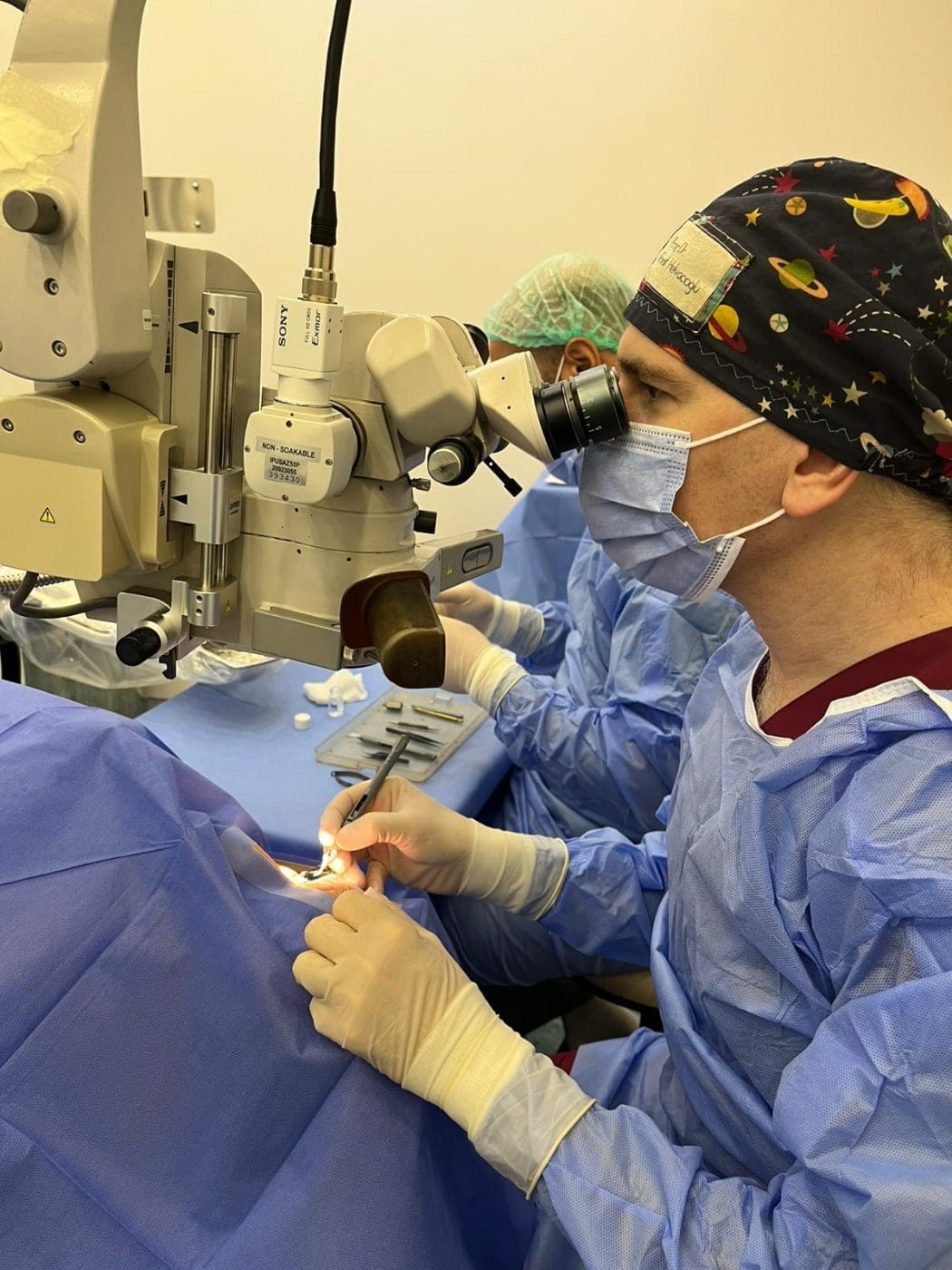
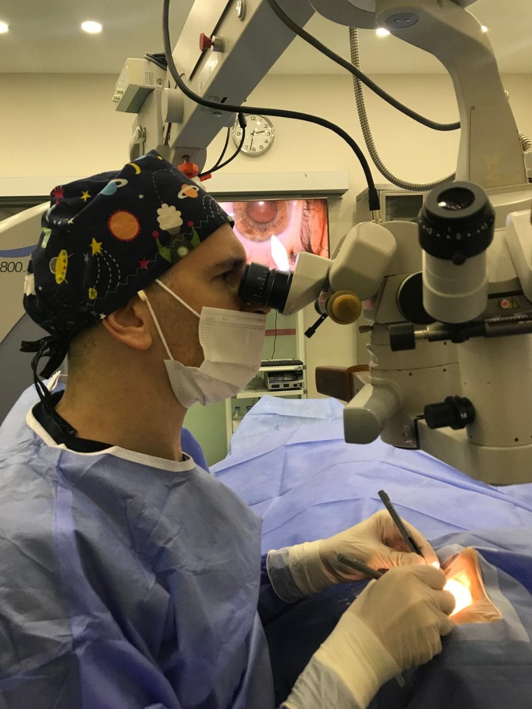
Today the standard technique is phacoemulsification (“phaco”). We perform the operation in an operating theatre under a surgical microscope and topical anaesthesia; you stay awake and feel no pain.
- Preparation and anaesthesia: First, drops dilate the pupil and anaesthetic drops numb the eye; then a sterile drape covers it.
- Micro-incision: The surgeon makes a self-sealing incision of about 2–2.8 mm at the edge of the clear front window of the eye (the cornea).
- Capsulorhexis: The surgeon then creates a circular opening in the front of the thin membrane (capsule) that surrounds the lens.
- Phacoemulsification: Ultrasound energy breaks the cloudy lens into small fragments, and the surgeon gently aspirates them from the eye.
- Lens implantation: Through the same incision, the surgeon inserts a foldable intraocular lens into the capsular bag, where it unfolds into position.
- Closure: The incision usually seals itself without stitches. Finally, a protective shield covers the eye.
The procedure usually takes 10–20 minutes. Patients can go home shortly afterwards, and the first follow-up examination takes place the next day.
Surgeon’s note — Prof. Dr. Fırat Helvacıoğlu
“In phacoemulsification the goal is to remove the lens while delivering as little energy to the eye as possible, because excessive ultrasound energy can stress the inner layer of the cornea. In studies published in international journals, we compared different ultrasound modes and phaco tip designs from exactly this perspective. We apply this experience in every operation, tailoring it to the density of each patient’s cataract.”
Which intraocular lens (IOL) options are available in cataract surgery?
Cataract surgery removes the natural lens and replaces it with an artificial lens that remains in the eye for life. The choice of lens determines at which distances you will see without glasses after surgery. We make the right choice together, based on your eye measurements, astigmatism, retinal and corneal health, occupation and expectations.
| Lens type | Distance seen clearly | Need for glasses | Suitable for |
|---|---|---|---|
| Monofocal | One distance (usually far) | Needed for near and often for intermediate distance | Patients who do not mind wearing glasses; some patients with retinal disease or glaucoma |
| Toric | Also corrects astigmatism | Depends on the lens type chosen | Patients with significant corneal astigmatism |
| EDOF (extended depth of focus) | Far and intermediate (computer, car dashboard) | You may need reading glasses for small print | Frequent night drivers; people who work a lot at intermediate distance |
| Trifocal (smart lens) | Far, intermediate and near | Not needed in daily life for most patients | Patients with otherwise healthy eyes who want independence from glasses |
What is a monofocal lens, and who is it suitable for?
A monofocal (single-focus) intraocular lens is the standard cataract lens that focuses light at one distance. Usually, we target distance vision, so you see far objects clearly without glasses. However, you need glasses for reading and often for computer distance. Because a monofocal lens does not split light into several foci, contrast sensitivity is high, and halos and glare at night are less common than with smart lenses.
- Patients who do not mind wearing glasses and value the highest possible image quality
- Patients with retinal or optic nerve disease such as macular degeneration, diabetic retinopathy or advanced glaucoma; we generally do not recommend multifocal lenses in these eyes
- People who drive a lot at night or are sensitive to halos
- Eyes that are not suitable for a smart lens because of corneal irregularity
Monovision and other monofocal lens options
Monovision with monofocal lenses: You can also reduce your dependence on glasses with monofocal lenses. We set the dominant eye for distance and the other eye for near–intermediate vision, leaving it slightly myopic. People who have previously used monovision with contact lenses usually adapt easily, and we can test suitability before surgery. Literature reviews show that pseudophakic monovision can provide a high rate of spectacle independence without a marked increase in photic phenomena.
Enhanced monofocal lenses: Their design aims to preserve monofocal image quality while providing somewhat better intermediate vision (computer, car dashboard). A meta-analysis of randomised trials showed a small but significant improvement in intermediate vision compared with standard monofocal lenses, with similar safety. However, you will usually still need reading glasses for near vision.
In patients with astigmatism, the toric version of a monofocal lens can correct astigmatism as well. According to a Cochrane review, multifocal lenses improve near vision and spectacle independence compared with monofocal lenses, but they also increase halos and glare. Therefore, we choose the lens according to each person’s priorities. Premium lenses (toric, EDOF, trifocal) cost more than a standard monofocal lens (see Cost and international patients).
More information: Smart lenses · Trifocal lens · EDOF lens · Toric lens
Technology and measurements before cataract surgery
Successful cataract surgery begins with the measurements taken before you enter the operating theatre. Optical biometry measures the length of the eye and the corneal curvature. We also assess corneal topography and thickness and examine the retina and macula with OCT. If you have dry eye, we treat it before surgery because it can distort the measurements.
Operations take place at Veni Vidi Eye Clinic, Caddebostan, with advanced phacoemulsification systems and surgical microscopes. We calculate the lens power with up-to-date formulas suited to your eye; for patients who have previously had laser eye surgery, we use dedicated calculation methods.
Cataract surgery after previous laser eye surgery
In eyes that have undergone laser vision correction for myopia, hyperopia or astigmatism (such as LASIK, PRK or SMILE), the shape of the cornea has changed. Therefore, calculating the intraocular lens power with standard formulas can be misleading. In these patients, we use calculation methods developed specifically for post-refractive eyes, together with additional measurements. If you have your prescription and records from before your laser treatment, bringing them to the consultation improves the accuracy of the calculation.
Will I see halos or shadows after cataract surgery? (Post-surgery dysphotopsia)
In the first weeks after surgery, some patients notice rings around lights, glare, or a faint shadow at the edge of their field of vision. The medical term for this is dysphotopsia. In most patients it fades or disappears within weeks to months as the brain adapts to the new lens. For the small number of cases in which it persists and is bothersome, treatment options are available.
There are two types
Positive dysphotopsia
Halos, glare, light streaks or starbursts around lights, noticed especially with headlights and street lamps at night.
Negative dysphotopsia
A thin, dark, crescent-shaped shadow on the temporal side of the visual field. It usually diminishes over time.
How common is it, and how long does it last?
According to the scientific literature, a considerable proportion of patients notice some light phenomena immediately after surgery. However, the proportion with persistent complaints after one year is low, and patients very rarely need surgical intervention.
| Condition | Early period | Persisting at 1 year |
|---|---|---|
| Positive dysphotopsia (halos, glare) | Up to 67% of patients | About 2% |
| Negative dysphotopsia (peripheral shadow) | Up to 26% | 0.13–3% |
Source: Pusnik et al., Life (Basel), 2023 — review.
In a multifocal (smart) lens study co-authored by Prof. Dr. Fırat Helvacıoğlu and published in the Turkish Journal of Ophthalmology in 2012, 40.7% of patients reported moderate halo/glare at one month. At three months, however, 74% reported no or only very mild complaints. By six months, glare had resolved, and halos were noticeable only when patients specifically looked for them. All patients in the study described themselves as “very satisfied” with the outcome.
What we do before surgery
- We choose the lens type according to your eye measurements and lifestyle (for example, frequent night driving).
- We evaluate pupil size and measurements related to the visual axis.
- We treat dry eye, if present, before surgery.
- We openly discuss possible light phenomena and the adaptation period before surgery.
What we do after surgery
- We monitor the adaptation (neuroadaptation) process at follow-up visits.
- We treat dry eye and any small residual refractive error.
- We open any secondary cataract (posterior capsule opacification) that develops later with a YAG laser.
- In the rare cases where complaints are persistent and bothersome, we consider surgical options such as repositioning or exchanging the lens.
Is cataract surgery risky?
Cataract surgery is one of the most common and most successful operations in the world, and vision improves markedly in the vast majority of patients. As with any surgery, there are some risks; if we recognise them early, we can treat most of them:
- Temporary rise in eye pressure or corneal oedema: Usually resolves with drops within a few days.
- Secondary cataract (posterior capsule opacification): Vision becomes cloudy again months or years later; a YAG laser procedure lasting a few minutes clears it. No pain is felt during the procedure.
- Intraocular infection (endophthalmitis): Very rare; sterile conditions and antibiotic prophylaxis minimise the risk.
- Retinal detachment and macular oedema: Uncommon; regular follow-up is especially important in highly myopic patients.
Secondary cataract (posterior capsule opacification) and YAG laser
During cataract surgery, the surgeon removes the contents of the cloudy natural lens and places the intraocular lens inside the lens’s thin membrane (the capsule). In some patients this membrane behind the lens becomes cloudy months or years later. Doctors call this secondary cataract or posterior capsule opacification (PCO). The cataract itself does not come back; what becomes cloudy is the membrane that holds the lens.
Symptoms
- Vision that had improved after surgery gradually becoming blurry again
- Glare in bright light and scattering of headlights at night
- Faded colours and contrast, difficulty reading
Treatment with YAG laser
- Preparation: Drops dilate the pupil and numb the eye.
- Laser: An Nd:YAG laser creates a small opening in the centre of the cloudy membrane. There is no incision and no need for stitches.
- Duration: The procedure takes a few minutes; thanks to the anaesthetic drops, most patients feel no pain. Patients usually return to daily life the same day.
- Follow-up: We plan drops and a check-up for a possible short-term rise in eye pressure.
The opening does not close again, so you usually need the procedure only once. Because a short-term rise in eye pressure, floaters and, very rarely, a retinal tear or detachment can occur, do not miss the follow-up examinations after the procedure.
If your vision becomes blurry again after surgery, it is important to have an examination; the cause may be something other than the capsule, such as dry eye or macular oedema.
Recovery after cataract surgery
| Time | What to expect | What to watch for |
|---|---|---|
| Day of surgery | Mild blurring, gritty sensation | Protective shield stays on; rest |
| Day 1 | First check-up; vision clears noticeably in most patients | Start the drops; do not rub the eye |
| Week 1 | Return to daily activities, reading and screen use | Keep water out of the eye; wear the shield at night |
| Weeks 2–4 | Vision sharpens and light adaptation improves | Avoid swimming pools, the sea and dusty environments |
| Months 1–3 | Vision stabilises; we prescribe glasses if needed | Attend follow-up visits regularly |
We advise returning to driving after we have checked your visual acuity at a follow-up visit. If both eyes have cataract, we usually operate on the second eye a few days to a few weeks later.
Cost of cataract surgery and information for international patients
The cost of cataract surgery varies from person to person, and we finalise it after the examination. The main factors are:
- The type of intraocular lens chosen (monofocal, toric, EDOF, trifocal)
- Whether you have surgery on one or both eyes
- The density of the cataract and whether there are other eye conditions
- Your health insurance cover (international or private insurance)
For international patients: Send us your previous eye reports by WhatsApp to receive a personalised treatment plan and quote. You can usually complete surgery for one eye, including the pre-operative examination and the next-day check, within a few days. If both eyes need surgery, we recommend planning to stay in Istanbul for about one week. Later, you can continue your follow-up with your own ophthalmologist, and consultations in English are available.
Why choose Prof. Dr. Fırat Helvacıoğlu for cataract surgery?
Prof. Dr. Fırat Helvacıoğlu is a cataract and refractive surgeon who has practised as an ophthalmologist since 2007. He continues his academic work as Head of the Department of Ophthalmology at Üsküdar University Faculty of Medicine and sees patients as a senior consultant in the Cataract and Refractive Surgery department of Veni Vidi Eye Clinic, Caddebostan.
- FEBOS-CR: One of the first ophthalmologists to receive the European Board of Ophthalmology subspecialty diploma in cataract and refractive surgery
- Scientific publications: 28 publications in Web of Science (h-index 8), including first-author cataract surgery papers in the Journal of Cataract & Refractive Surgery and the American Journal of Ophthalmology
- Peer review: Reviewer for Ophthalmology (AAO), the Journal of Cataract & Refractive Surgery and the International Journal of Ophthalmology; member of the IJO International Editorial Board
- Lens calculation: Member of the international study group of the new-generation Hoffer H5 biometry formula; his “Helvacıoğlu coefficient optimizing factor” for torsional phacoemulsification was presented at the 36th ESCRS Congress (Vienna, 2018)
- International qualifications: ICO Basic Science (2007) and Clinical Science (2009) examinations
- Memberships: Turkish Ophthalmological Association (active member of the Cataract and Refractive Surgery Unit), ESCRS, ASCRS
- Clinical decision support: Member of the Medical Advisory Board of the RLES AI platform, developed for cataract and refractive surgery planning
Frequently asked questions about cataract surgery
No. We use topical (eye-drop) anaesthesia, which numbs the eye completely. You may feel some pressure during surgery, but no pain. A short-lived gritty sensation afterwards is normal.
Usually not. Topical anaesthesia is sufficient and the patient stays awake. If needed, we can give mild sedation; in special cases, we plan other anaesthetic methods.
The operation usually takes 10–20 minutes. Including preparation and a short observation period afterwards, you spend a few hours at the clinic and go home the same day.
No. The removed natural lens does not grow back. However, in some patients the membrane behind the lens becomes cloudy months or years later (secondary cataract); a YAG laser procedure lasting a few minutes clears it.
Yes. Intraocular lenses are permanent, need no replacement or maintenance, and under normal circumstances remain in the eye for life.
It depends on the type of lens implanted. With a monofocal lens, you will usually need reading glasses for near vision; with trifocal (smart) lenses, most patients see without glasses in daily life. We evaluate the options together during your consultation.
Usually, we operate on each eye on a separate day, a few days to a few weeks apart. This allows us to see how the first eye heals and to plan the second eye accordingly.
Yes. You can have cataract surgery in any season. In summer you only need to avoid swimming pools and the sea during the first weeks, wear sunglasses and protect the eye from dust and sweat.
This depends on your visual acuity and your follow-up examination. Many patients can return to driving within a few days, but this decision should come after the check-up.
References
- Pusnik A, Petrovski G, Lumi X. Dysphotopsias or Unwanted Visual Phenomena after Cataract Surgery. Life (Basel). 2023;13(1):53. doi:10.3390/life13010053
- Masket S, Fram NR. Pseudophakic Dysphotopsia: Review of Incidence, Cause, and Treatment of Positive and Negative Dysphotopsia. Ophthalmology. 2021;128(11):e195–e205. doi:10.1016/j.ophtha.2020.08.009
- Tunç Z, Helvacıoğlu F, Şencan S, Şimşek Ş. Multifocal progressive refractive-diffractive lens: optical design and clinical results. Turkish Journal of Ophthalmology. 2012. Article (in Turkish)
- Helvacıoğlu F et al. Comparison of two different ultrasound methods of phacoemulsification. American Journal of Ophthalmology. 2014. doi:10.1016/j.ajo.2014.04.015
- Helvacıoğlu F et al. Torsional phacoemulsification with 30-degree and 45-degree aperture tips. Journal of Cataract & Refractive Surgery. 2014. doi:10.1016/j.jcrs.2013.07.051
- Helvacıoğlu F et al. Torsional phacoemulsification with 12-degree and 22-degree bent tips. Journal of Cataract & Refractive Surgery. 2013. doi:10.1016/j.jcrs.2013.03.002
- Wanniarachchi K, Mehta JS. Management of positive and negative dysphotopsia postcataract surgery – A literature review. Taiwan J Ophthalmol. 2025;15(4):572–579. doi:10.4103/tjo.TJO-D-25-00017
- de Silva SR et al. Multifocal versus monofocal intraocular lenses after cataract extraction. Cochrane Database Syst Rev. 2016;12(12):CD003169. doi:10.1002/14651858.CD003169.pub4
- Labiris G et al. A systematic review of pseudophakic monovision for presbyopia correction. Int J Ophthalmol. 2017;10(6):992–1000. doi:10.18240/ijo.2017.06.24
- Loh NC et al. Enhanced monofocal versus conventional monofocal intraocular lenses: a systematic review and meta-analysis of randomized controlled trials. Can J Ophthalmol. 2026;61(1):12–19. doi:10.1016/j.jcjo.2025.07.010
- Tetikoğlu M, Yeter C, Helvacıoğlu F, Aktaş S, Sağdık HM, Özcura F. Effect of Corneal Incision Enlargement on Surgically Induced Astigmatism in Biaxial Microincision Cataract Surgery. Türk Oftalmoloji Dergisi. 2016;46(3):99-103. doi:10.4274/tjo.52386
- Helvacıoğlu F, Şencan S, Tunç Z, Yeter C. Torsiyonel Fakoemülsifikasyon ve Fako İğnesi Seçimi. Türk Oftalmoloji Dergisi. 2014;44(5):392-395. doi:10.4274/tjo.59389
- Helvacioglu F, Tunc Z, Yeter C, Oguzhan H, Sencan S. Ozil IP torsional mode versus combined torsional/longitudinal microcoaxial phacoemulsification. European Journal of Ophthalmology. 2012;22(6):936-942. doi:10.5301/ejo.5000136
- Yuzbasioglu E, Helvacioglu F, Tugcu B, Terzi N, Keskinbora K. The results of cataract nigra cases operated with the mini-nuc technique. International Ophthalmology. 2009;29(6):451-457. doi:10.1007/s10792-008-9265-8
This page is for patient information and does not replace an examination or medical assessment. Your doctor decides on the right treatment for you after an eye examination.
Book a cataract consultation
Prof. Dr. Fırat Helvacıoğlu · Veni Vidi Eye Clinic, Bağdat Caddesi No: 257, Caddebostan – Kadıköy, Istanbul, Türkiye