
Keratoconus Treatment
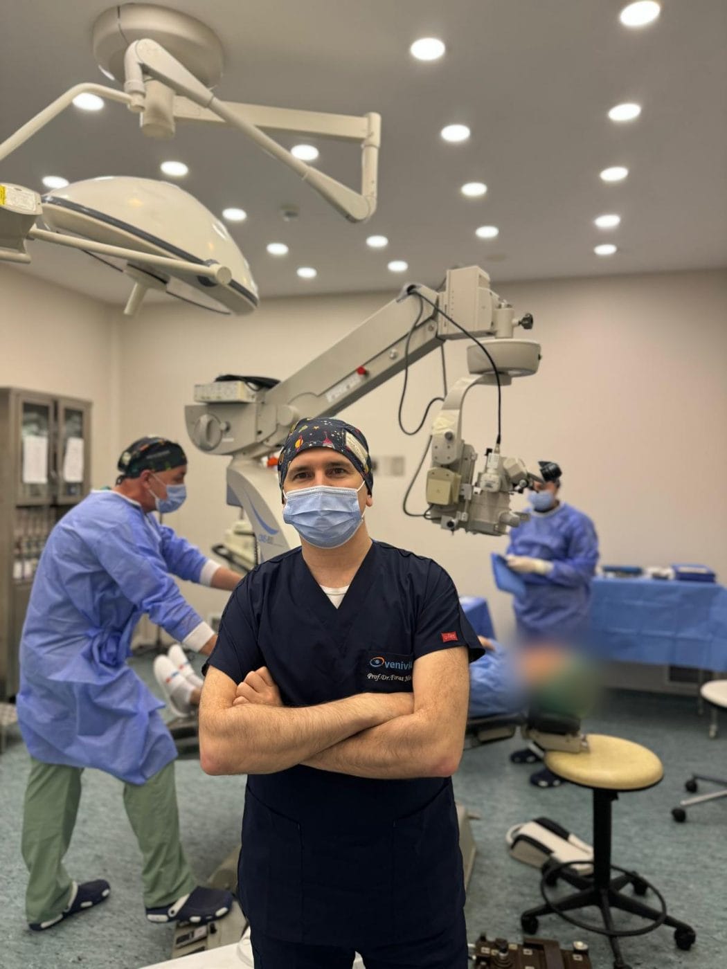
Cross-linking (CXL) · Combined CXL + No Touch · Corneal ring segments and CAIRS · Prof. Dr. Fırat Helvacıoğlu, FEBOS-CR · Veni Vidi Eye Clinic, Caddebostan – Kadıköy, Istanbul
Keratoconus is a progressive disease in which the cornea thins and bulges into a cone shape. In keratoconus treatment, the first step is to halt progression with corneal cross-linking (CXL). Then, to improve vision, the options include combined CXL + No Touch laser, CAIRS and intracorneal ring segments (ICRS), specialty contact lenses or an implantable lens. Early diagnosis largely prevents the need for a corneal transplant.
What is keratoconus?
Keratoconus is a progressive corneal disease in which the cornea — the clear front surface of the eye — gradually thins and bulges forward into a cone shape. As the cornea loses its regular shape, irregular astigmatism develops, and glasses can no longer fully correct vision. It usually affects both eyes, although one eye may be more advanced than the other.
Keratoconus is more common than generally thought: a nationwide registry study in the Netherlands reported a prevalence of about 1 in 375 people, with a mean age at diagnosis of 28. The disease usually begins in adolescence and may progress into the 30s and 40s; therefore, early diagnosis is the key to preserving vision.
What are the symptoms of keratoconus?
- Frequently changing prescription: Especially an increase in astigmatism at short intervals.
- Vision not fully corrected with glasses: Inability to see clearly despite new glasses.
- Blurred, shadowed or double vision: Letters may appear doubled even when looking with one eye.
- Light sensitivity and night vision problems: Scattering and glare around headlights.
- Contact lenses becoming uncomfortable: The lens slipping or not staying in place.
If you have these symptoms, particularly in adolescence and early adulthood, you should have a detailed corneal screening. However, in the early stage, vision is often normal, and only corneal topography can detect the disease.
What causes keratoconus? Risk factors
The exact cause of keratoconus is unknown; however, experts believe that a combination of genetic predisposition and environmental factors plays a role.
- Family history: The risk is higher in people with a first-degree relative with keratoconus; therefore, we recommend screening family members.
- Eye rubbing: This is the modifiable risk factor with the strongest link to the development and progression of keratoconus. If your eyes itch, you should treat the itch rather than rub your eyes.
- Allergy and atopy: Large population studies have shown associations between keratoconus and allergic diseases, asthma and some immune-mediated diseases.
How do we diagnose keratoconus?
We diagnose keratoconus with corneal topography and tomography, which map the front and back surfaces of the cornea, a corneal thickness map (pachymetry) and a detailed eye examination. To determine whether the disease is progressing, we repeat the measurements at regular intervals and compare them. The Global Consensus on Keratoconus also recommends evaluating changes in the anterior and posterior corneal surfaces and thickness together to document progression.
Comprehensive screening before laser eye surgery is also vital for detecting early (subclinical) keratoconus that causes no symptoms: conventional laser treatment of a keratoconic cornea can accelerate the disease. Prof. Dr. Fırat Helvacıoğlu is a member of the Medical Advisory Board of RLES AI, a clinical decision support platform. He has also contributed to its keratoconus screening module and to its module for distinguishing temporary contact lens–induced corneal changes.
Keratoconus treatment: which method suits whom?
Keratoconus treatment has two goals: halting disease progression and improving visual quality. We plan keratoconus treatment individually according to the stage of the disease, whether it is progressing, corneal thickness and age.
| Treatment | Goal | Suitable for |
|---|---|---|
| Glasses / soft lenses | Correct vision | Very early stage, eyes with little irregularity |
| Specialty contact lenses (RGP, hybrid, scleral) | Neutralise the optical effect of the irregular cornea | Any stage in which glasses do not provide adequate vision |
| Cross-linking (CXL) | Halt progression | Progressive keratoconus, especially in children and young people |
| Combined CXL + No Touch | Halt progression and reduce irregularity | Mild to moderate stage, adequate corneal thickness, patients who cannot wear lenses |
| CAIRS and intracorneal ring segments (ICRS) | Flatten the cone and improve vision | Moderate stage, clear central cornea, poor lens tolerance |
| Implantable lens (ICL) | Correct the remaining prescription | Selected patients whose disease has stabilised |
| Corneal transplantation (DALK / PK) | Restore vision | Advanced stage, corneal scarring, or failure of other methods |
Today, thanks to early diagnosis and cross-linking, keratoconus treatment is possible without the need for a corneal transplant in the great majority of patients.
Contact lenses in keratoconus treatment
Contact lenses are the most effective non-surgical way to correct vision in keratoconus. Because the surface of a keratoconic cornea is irregular, glasses cannot fully correct this irregularity. Specialty lenses, thanks to the tear layer between the eye and the lens, create a new, regular optical surface in front of the cornea. As a result, many patients who cannot see adequately with glasses see much more clearly with a suitable lens.
| Lens type | Features | Suitable for |
|---|---|---|
| Soft toric / keratoconus soft lens | Comfortable; specially designed thick-centre models can mask mild irregularity | Early stage, eyes with little irregularity |
| Rigid gas-permeable (RGP) lens | Rests on the cornea; the classic option and one of the most effective at correcting an irregular surface. Keratoconus-specific designs (e.g. multi-curve lenses) are available | Mild to moderate stage; requires an adaptation period |
| Hybrid lens | Rigid gas-permeable centre with a soft silicone hydrogel skirt; combines the visual quality of a rigid lens with the comfort of a soft lens | Mild to moderate (and selected advanced) cases who cannot adapt to rigid lenses or whose rigid lenses decentre; particularly central cones |
| Piggyback (dual lens) | A rigid lens worn over a soft lens | Patients who cannot tolerate a rigid lens alone |
| Scleral / mini-scleral lens | A large-diameter lens that rests on the white of the eye (sclera) without touching the cornea; the fluid reservoir beneath protects the cornea and neutralises irregularity | Moderate to advanced stage, steep cones, after ring segments or transplantation, patients who cannot tolerate rigid lenses |
Hybrid lenses in keratoconus
A hybrid lens is a contact lens with a rigid gas-permeable centre and a soft silicone hydrogel “skirt”. Together with the tear layer beneath it, the rigid centre creates a regular optical surface in front of the irregular cornea, providing rigid-lens-quality vision. The soft skirt helps centre the lens on the eye, reduces movement and dislodgement, and offers more comfortable wear than a rigid lens. In addition, manufacturers design new-generation hybrid lenses to vault gently over the cone without pressing on the cornea.
Hybrid lenses: who they suit and what studies show
- Who is it suitable for? Patients who cannot adapt to rigid (RGP) lenses, whose rigid lenses decentre or fall out, who play sports, and who cannot see adequately with soft lenses. Success rates are particularly high with centrally located cones.
- Vision: A comparative study from Türkiye showed that hybrid lenses provide visual gains equivalent to rigid lenses in moderate and advanced keratoconus. In another study of 110 keratoconic eyes, mean visual acuity improved from 0.36 with spectacles to 0.80 (decimal) with hybrid lenses; in most patients, the first one or two trials were enough to find the right lens.
- Quality of life: In patients unable to tolerate rigid or soft lenses, studies report that new-generation hybrid lenses markedly improve vision-related quality of life scores.
- After cross-linking: In patients using hybrid lenses after CXL, vision improved and lens wear did not adversely affect disease stabilisation.
- Points to consider: Inserting and removing a hybrid lens requires practice; lens life is shorter and the cost higher than with soft lenses. In one study, about one third of patients discontinued the lens within the first 6 months, mostly because of comfort and handling difficulties; success depends on the shape and location of the cone. For inferior and peripheral cones, however, scleral lenses may be more suitable.
| Feature | Hybrid lens | Scleral lens |
|---|---|---|
| Where it rests | The cornea and its immediate surroundings | The white of the eye (sclera), without touching the cornea |
| Best suited cone | Central, mild to moderate cones | Steep, advanced, inferior or peripheral cones |
| Insertion and removal | Similar to a soft lens; requires practice | Inserted after filling the bowl with a special solution; longer learning curve |
| Comfort | Better than a rigid lens | Usually the most comfortable option, as there is no corneal contact |
Scleral lenses and practical advice on lens wear
Scleral lenses can be an alternative to corneal transplantation, particularly in advanced keratoconus. For example, in a study of 89 keratoconic eyes, mini-scleral lenses significantly improved visual acuity and vision-related quality of life; the most common reason for discontinuation was difficulty with insertion and removal. Even in advanced cases with a history of corneal hydrops, most suitable patients could achieve functional vision with scleral lenses. Therefore, researchers recommend trying scleral lenses before deciding on a transplant.
After cross-linking, combined treatment, CAIRS or intracorneal ring segments (ICRS), the shape of the cornea changes, so we need to reassess the lens choice; after these treatments, lenses fit more comfortably in many patients.
How do we choose the right lens?
At the consultation, Prof. Dr. Fırat Helvacıoğlu determines and recommends the most suitable lens type for you based on corneal tomography, the location and stage of the cone, your tear film and your daily habits. You then obtain the lens according to this recommendation, and we monitor your corneal health at follow-up visits once you start wearing it.
Prof. Dr. Fırat Helvacıoğlu’s experience in keratoconus treatment
Surgeon’s experience and publications
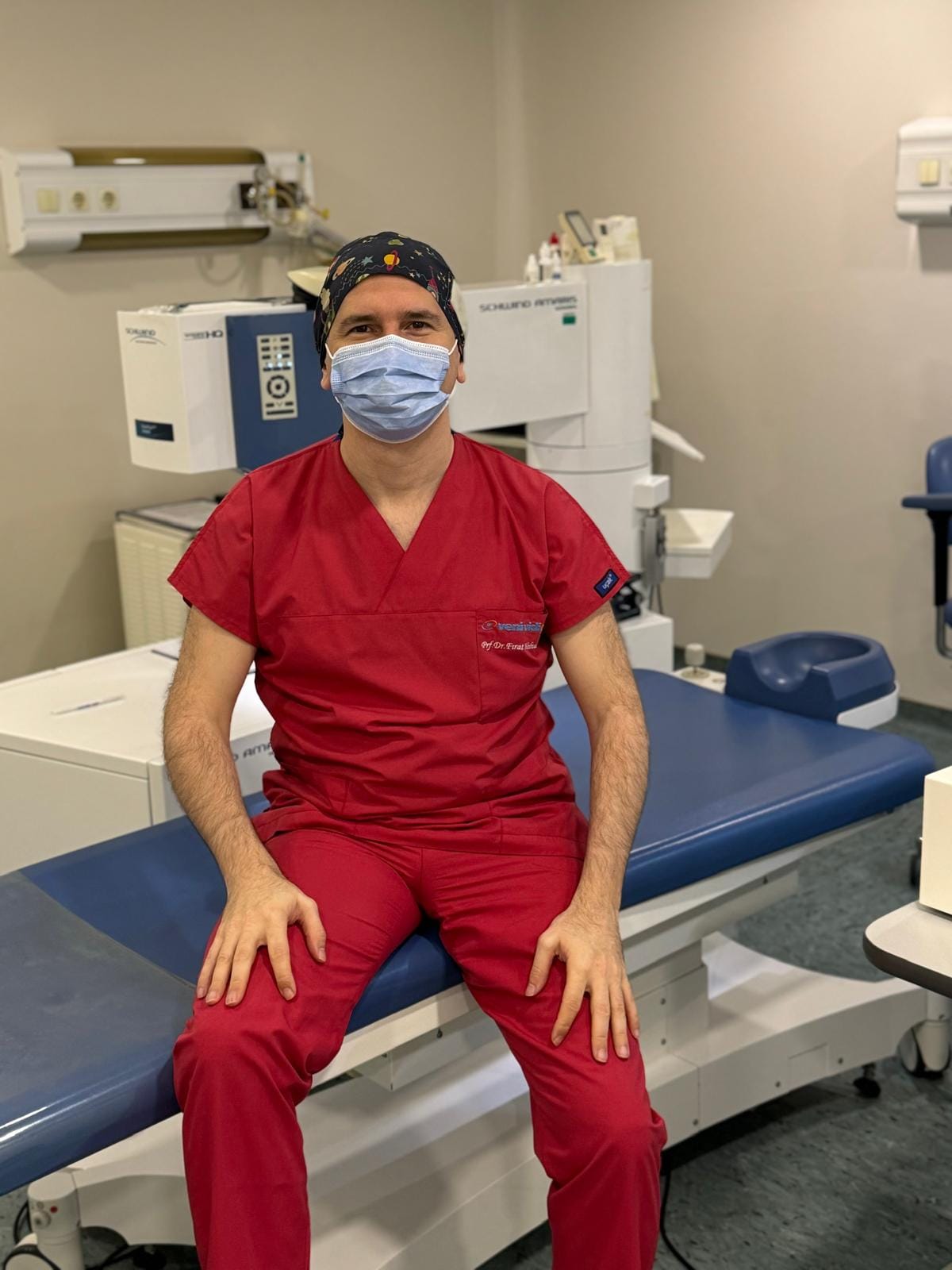
Prof. Dr. Fırat Helvacıoğlu is a co-author of two clinical studies on intracorneal ring segment treatment. In 30 keratoconic eyes, the surgeons implanted the rings with a special mechanical dissection technique. At 1 year, the mean spherical equivalent decreased from −6.42 D to −1.26 D and mean corneal steepness from 49.4 D to 44.4 D, with no serious complications. A study of 12 eyes with post-LASIK ectasia likewise reported significant improvement.
He has also presented the short-term, 2-year, 3-year and long-term results of a hybrid treatment combining corneal wavefront-guided TransPRK with cross-linking in the same session in keratoconus at ESCRS congresses between 2016 and 2019.
Keratoconus treatment in children and young people
Keratoconus tends to progress faster in children and young people. In the randomised KERALINK trial in the United Kingdom, involving patients aged 10–16, cross-linking reduced the likelihood of progression by about 90% compared to standard monitoring. As a result, the researchers recommended considering CXL as first-line treatment for progressive keratoconus. Young patients diagnosed with keratoconus therefore need close follow-up and treatment without delay when we detect progression.
Cost of keratoconus treatment and information for international patients
We determine the cost of keratoconus treatment after the examination, according to the planned procedure (CXL, combined treatment, CAIRS or intracorneal ring segments/ICRS) and whether it involves one or both eyes. For international patients: send us your corneal topography/tomography and previous reports via WhatsApp for a preliminary assessment and quote. We can plan most keratoconus treatments so that you complete the treatment and early follow-up within about one week in Istanbul; later follow-up can then continue with your own ophthalmologist. Consultations are available in English.
Frequently asked questions about keratoconus and its treatment
Keratoconus does not usually cause complete blindness. However, without treatment, vision can decrease severely, and the advanced stage may require a corneal transplant. Early diagnosis and cross-linking can halt progression in the great majority of patients.
Surgeons do not perform conventional laser eye surgery (such as LASIK) on keratoconic eyes, because it can accelerate the disease. However, in selected mild to moderate cases, we can combine a personalised, low-dose No Touch laser treatment with cross-linking, which strengthens the cornea.
The disease usually begins in adolescence and may progress into the 30s and 40s; the rate of progression varies from person to person. You should therefore have regular measurements regardless of age.
Genetic predisposition plays an important role. Therefore, we advise the parents, siblings and children of someone with keratoconus to undergo corneal screening.
Yes. Soft lenses in the early stage, and rigid (RGP) or scleral lenses in more advanced stages, can markedly improve vision. However, lenses do not halt progression; if the disease is progressing, cross-linking is necessary.
There is no single best lens. Instead, specialty soft lenses stand out in the early stage, rigid (RGP) or hybrid lenses in mild to moderate stages, and scleral lenses in advanced stages and steep cones. Your doctor decides which lens is suitable at the consultation, based on corneal tomography, your tear film and your daily wearing habits.
A hybrid lens is a contact lens with a rigid gas-permeable centre and a soft outer part. It combines rigid-lens-quality vision with comfort close to that of a soft lens. It is a good option for patients with mild to moderate keratoconus who cannot adapt to rigid lenses, whose rigid lenses decentre or fall out, and especially those with central cones.
Hybrid lenses are generally more successful for central, mild to moderate cones, while scleral lenses are generally more successful for steep, advanced or inferior cones. Your doctor decides which is suitable at the consultation, based on corneal tomography.
A scleral lens is a large-diameter rigid lens that rests on the white of the eye without touching the cornea. The fluid layer beneath it neutralises corneal irregularity and protects the cornea. It is an option in advanced keratoconus, in patients who cannot tolerate rigid lenses, and in irregular corneas after ring segments or transplantation.
We perform the procedures under topical anaesthesia, so you feel no pain during them. After cross-linking and combined treatments that remove the surface layer, grittiness, watering and light sensitivity may occur for the first 2–3 days.
References
- Godefrooij DA et al. Age-specific incidence and prevalence of keratoconus: a nationwide registration study. Am J Ophthalmol. 2017;175:169–172. doi:10.1016/j.ajo.2016.12.015
- Gomes JA et al. Global consensus on keratoconus and ectatic diseases. Cornea. 2015;34(4):359–369. doi:10.1097/ICO.0000000000000408
- Larkin DFP et al. Effect of corneal cross-linking versus standard care on keratoconus progression in young patients: the KERALINK randomized controlled trial. Ophthalmology. 2021;128(11):1516–1526. doi:10.1016/j.ophtha.2021.04.019
- Claessens JLJ et al. Nationwide epidemiological approach to identify associations between keratoconus and immune-mediated diseases. Br J Ophthalmol. 2022;106:1350–1354. doi:10.1136/bjophthalmol-2021-318804
- Tunc Z, Helvacioglu F, Sencan S. Evaluation of intrastromal corneal ring segments for treatment of keratoconus with a mechanical implantation technique. Indian J Ophthalmol. 2013;61(5):218–225. doi:10.4103/0301-4738.109519
- Tunc Z, Helvacioglu F, Sencan S. Evaluation of intrastromal corneal ring segments for treatment of post-LASIK ectasia patients with a mechanical implantation technique. Indian J Ophthalmol. 2011;59(6):437–443. doi:10.4103/0301-4738.86310
- Shajari M et al. Comparison of standard and accelerated corneal cross-linking for the treatment of keratoconus: a meta-analysis. Acta Ophthalmol. 2019;97(1):e22–e35. doi:10.1111/aos.13814
- Taşcı YY et al. Comparison of hybrid contact lenses and rigid gas-permeable contact lenses in moderate and advanced keratoconus. Turk J Ophthalmol. 2023;53(3):142–148. doi:10.4274/tjo.galenos.2022.82754
- Serdarov A et al. Clinical performance and patient satisfaction of hybrid contact lenses in patients with keratoconus. Turk J Ophthalmol. 2023;53(4):206–212. doi:10.4274/tjo.galenos.2022.43077
- Kloeck D, Koppen C, Kreps EO. Clinical outcome of hybrid contact lenses in keratoconus. Eye Contact Lens. 2021;47(5):283–287. doi:10.1097/ICL.0000000000000738
- Ozcan DO, Ozcan SC. Effects of a new-generation hybrid contact lens on visual performance and vision-related quality of life in patients with keratoconus. Arq Bras Oftalmol. 2023;86(1):7–12. doi:10.5935/0004-2749.20230001
- Aslan MG et al. The impact of hybrid contact lenses on keratoconus progression after accelerated transepithelial corneal cross-linking. Int Ophthalmol. 2021;41(1):45–55. doi:10.1007/s10792-020-01551-w
- Kreps EO et al. Mini-scleral lenses improve vision-related quality of life in keratoconus. Cornea. 2021;40(7):859–864. doi:10.1097/ICO.0000000000002518
- Kreps EO, Claerhout I, Koppen C. The outcome of scleral lens fitting for keratoconus with resolved corneal hydrops. Cornea. 2019;38(7):855–858. doi:10.1097/ICO.0000000000001946
- Swartz G et al. Impact of contact lens correction on wavefront aberrations and vision quality in keratoconus. Ophthalmic Physiol Opt. 2025;45(7):1811–1828. doi:10.1111/opo.70037
- Deboutte I et al. Influence of specialty contact lens wear on posterior corneal tomography in keratoconus subjects. Eye Contact Lens. 2022;48(12):497–502. doi:10.1097/ICL.0000000000000941
This page provides information for patients and does not replace an examination or medical assessment. We determine the keratoconus treatment that is right for you after an eye examination and measurements.
Book a keratoconus consultation and corneal screening
Prof. Dr. Fırat Helvacıoğlu · Veni Vidi Eye Clinic, Bağdat Caddesi No: 257, Caddebostan – Kadıköy, Istanbul, Türkiye