Pterygium and Pinguecula
Pterygium surgery with conjunctival autograft · Mitomycin C · Prof. Dr. Fırat Helvacıoğlu, FEBOS-CR · Veni Vidi Eye Clinic, Caddebostan – Kadıköy, Istanbul
A pterygium is a triangular growth of the membrane covering the white of the eye onto the cornea. A pinguecula, in contrast, is a yellowish patch that does not extend onto the cornea. The main cause is sunlight. Pterygium surgery with a conjunctival autograft is the standard treatment for a pterygium that affects vision or is progressing. In this operation, the surgeon removes the tissue and covers the area with the patient’s own conjunctiva. As a result, the risk of recurrence falls markedly.
What are pterygium and pinguecula?
A pterygium is a triangular, vascular growth of the conjunctiva — the membrane covering the white of the eye — onto the cornea. It usually starts on the nasal side. A pinguecula is a yellowish, slightly raised area of degeneration on the white of the eye that does not extend onto the cornea.
The main cause of both is long-term exposure to sunlight (UV); wind, dust and dry climates also play a role. According to a meta-analysis of 415,911 people, the worldwide prevalence of pterygium is about 12%. In addition, more than 5 hours of sun exposure a day and working outdoors increase the risk, while wearing sunglasses roughly halves it.
Pterygium surgery: the surgical steps
Shows the appearance of pterygium and surgery with an autograft, step by step.
- PingueculaA yellowish, slightly raised patch on the white of the eye that does not extend onto the cornea; it relates to sun and wind exposure.
- PterygiumA triangular, vascular tissue extends from the white of the eye onto the cornea, causing grittiness, redness and astigmatism.
- Excision and graft planningFirst, the surgeon carefully removes the pterygium from the corneal surface. Then the surgeon prepares a piece of healthy conjunctiva (autograft) from the upper area.
- Closure with an autograftThe surgeon covers the bare area with the patient’s own conjunctiva (tissue glue or fine sutures); if needed, the surgeon uses mitomycin C to reduce the risk of recurrence.
Symptoms, and when do you need pterygium surgery?
- Redness, stinging, burning and a foreign-body sensation
- A raised, fleshy tissue on the eye; cosmetic concern
- Astigmatism and blurred vision caused by traction on the cornea
- In advanced cases, reaching the pupil area and obstructing vision
We recommend pterygium surgery in these situations: Progression towards the visual axis, increasing astigmatism and blurred vision, symptoms not relieved by drops, interference with contact lens wear, and cosmetic concern. In patients planning cataract or laser surgery, we usually treat the pterygium first because it can distort the measurements.
Treatment: from drops to pterygium surgery with an autograft
For small, asymptomatic pterygium and pinguecula, preservative-free artificial tears, short courses of anti-inflammatory drops during flare-ups and UV protection may be sufficient.
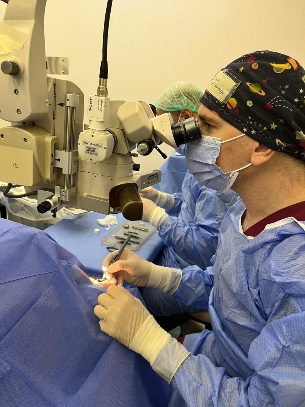
Surgery: The surgeon carefully removes the pterygium from the corneal surface under local anaesthesia. With the old technique, the surgeon simply excises the tissue and leaves the area bare. Then the recurrence rate can exceed 80%. Today the standard technique is to cover the bare area with the patient’s own healthy conjunctiva (conjunctival autograft). The surgeon secures the graft with tissue glue or fine sutures. According to a Cochrane review, a conjunctival autograft roughly halves the risk of recurrence compared with an amniotic membrane graft. In patients at high risk of recurrence, mitomycin C applied during or before surgery further reduces recurrence significantly.
Grittiness and redness may occur for a few days after surgery; you use antibiotic and corticosteroid drops for a few weeks. Sun protection is important to prevent recurrence.
Prof. Dr. Fırat Helvacıoğlu’s research on pterygium surgery
Research
Pterygium is one of the subjects Prof. Dr. Fırat Helvacıoğlu has studied most extensively. He presented the 1- and 2-year results of pre-operative subconjunctival mitomycin C and superior temporal limbal conjunctival autograft techniques for preventing pterygium recurrence at ESCRS (Lisbon 2005, London 2006), ASCRS (Washington 2005) and Mediterranean Ophthalmological Society congresses, and conducted studies comparing these two techniques. He has published and presented work on the surgical management of scleromalacia after mitomycin C (ESCRS Paris 2004; MN Oftalmoloji 2007), the long-term results of free conjunctival autografts and an extended limbal conjunctival autograft technique (ESCRS Vienna 2018). His study of the incidence of pterygium in a university hospital was published in the Journal of Clinical and Experimental Investigations (2013).
Prevention
- Outdoors, wear wraparound sunglasses with UV400 protection and a hat.
- Protect your eyes in windy and dusty environments; keep them moist with artificial tears.
- Take particular care to protect your eyes from the sun in the first months after surgery.
Frequently asked questions
No. A pterygium does not regress on its own; some remain stable for years while others advance towards the cornea. We recommend monitoring and, when necessary, surgery.
Pterygium surgery with an autograft usually takes 30–45 minutes under local anaesthesia, and you go home the same day.
With an autograft and, when needed, mitomycin C, we markedly reduce the risk of recurrence. However, recurrence can still occur, particularly in young patients with heavy sun exposure. Sun protection is important.
Usually not. We monitor it with drops and sun protection. Rarely, we may remove it if it becomes frequently inflamed or causes cosmetic concern.
It can cause astigmatism by pulling on the cornea, and can obstruct vision when it advances into the pupil area.
References
- Rezvan F et al. Prevalence and risk factors of pterygium: a systematic review and meta-analysis. Surv Ophthalmol. 2018;63(5):719–735. doi:10.1016/j.survophthal.2018.03.001
- Clearfield E et al. Conjunctival autograft for pterygium. Cochrane Database Syst Rev. 2016;2(2):CD011349. doi:10.1002/14651858.CD011349.pub2
- Zeng W et al. Anti-fibrotic, anti-VEGF or radiotherapy treatments as adjuvants for pterygium excision: a systematic review and network meta-analysis. BMC Ophthalmol. 2017;17(1):211. doi:10.1186/s12886-017-0601-5
- Turgut FG, Helvacıoğlu F. Incidence of pterygium in patients admitted to a university hospital. J Clin Exp Invest. 2013;4:436–442.
- Tuğcu B, Helvacıoğlu F, Yüzbaşıoğlu E, Ağaçhan A. Two-year results of pre-operative subconjunctival mitomycin C in the prevention of pterygium recurrence. Bakırköy Tıp Dergisi. 2009;5:18–21 (in Turkish).
This page offers patient information and does not replace an examination or medical assessment. We determine the right treatment for you after an eye examination and measurements.
Pterygium assessment
Prof. Dr. Fırat Helvacıoğlu · Veni Vidi Eye Clinic, Bağdat Caddesi No: 257, Caddebostan – Kadıköy, Istanbul, Türkiye
Related pages
- Corneal Diseases and TreatmentsCorneal diseases such as keratitis, Fuchs dystrophy and keratoconus, and corneal transplantation (PK, DALK, DMEK): diagnosis and treatment in Istanbul.
- Dry Eye Disease and TreatmentDry eye treatment for meibomian gland dysfunction: symptoms, causes, diagnosis, and care with preservative-free tears, lid hygiene and punctal plugs.
- Comprehensive Eye ExaminationWho needs an eye examination and how often, what a comprehensive eye exam involves, and which symptoms need urgent care. Veni Vidi Eye Clinic, Istanbul.
- Eye Diseases and TreatmentsGlaucoma, dry eye, blepharitis, pterygium, corneal diseases, tear duct obstruction, strabismus and eyelid surgery in Istanbul. Diagnosis and treatment.